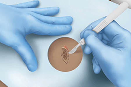
During an excisional biopsy, a scalpel is used to cut out a lump or an area of irregular skin and some surrounding healthy skin.
Before the mole is removed, your doctor will explain what they are going to do and will ask for permission (consent) to do the excision biopsy. The procedure usually takes place lying down. A series of questions are asked out loud to make sure there will be no problems during the operation. This includes questions to check there are no allergies and that everyone confirms which lesion is being removed.
The skin is cleaned with an antiseptic liquid and a clean sheet or drape is put over the skin. The skin is made numb using an injection of local anaesthetic. This part can be uncomfortable but is very quick. The procedure itself should not be painful in any way. If it is, more local anaesthetic will be given. Generally, stitches are needed to close the wound and a plaster put on the wound.
The tissue that is removed is sent to the laboratory. A different doctor examines it under a microscope. They will write a report on what they find, but this can take up to 3-4 weeks.
The stitches are removed after 5 to 14 days, depending on where the surgery was. If you need the sutures removed, then you will be given that appointment before you leave. Some stitches that dissolve and do not need to be removed.
The wound is not usually very sore afterwards. Any mild discomfort can be controlled with paracetamol.
Wound care advice will be given. Usually, the wound is covered and kept dry for 48 hours. After this time, the plaster is removed and brief showers are allowed (avoiding rubbing the wound with a towel after).
Increasing pain, redness or discharge is a sign of possible infection which would require medical attention.

Photodynamic therapy (PDT) destroys harmful cells, including cancer cells, using a combination of light and drugs called photosensitizers. Treatment happens in two parts. First, you’ll receive a photosensitizing drug. Then, a healthcare provider will expose the diseased cells to light. Light activates the photosensitizer, creating a chemical reaction that destroys the harmful cells.
Photodynamic therapy can only treat areas of your body where light can reach. This makes it especially useful for treating some skin cancers and precancers. Sometimes, providers use PDT to destroy cells on the lining of organs that they can reach with a light source.
Healthcare providers use PDT to treat cancers and pre-cancers eg superficial basal cell cancers, actinic keratosis and Bowen disease
.

Hyperhidrosis of the armpits is excessive sweating from the armpits which occurs because the sweat glands are over-active. Hyperhidrosis can also occur in other areas of the body, for example the hands and feet. Botulinum toxin (Botox) injections is a treatment that helps to control the symptoms of severe underarm sweating when other treatments have not been successful. Botulinum toxin is injected into the skin and works by temporarily blocking the chemical signals from the nerves that stimulate the sweat glands. The treatment will only work on the areas of the body that have been injected.
The benefit of this treatment is that it can reduce, and in most cases stop, the sweat produced in your armpits for up to 6 months. This can improve your confidence and social life as you no longer have to worry about the smell, sweat marks on your clothes or having to carry a spare top around with you. Most people find that they can stop using deodorants completely whilst having this treatment.
Botulinum toxin (Botox) injections is a treatment that helps to control the symptoms of severe underarm sweating when other treatments have not been successful. Botulinum toxin is injected into the skin and works by temporarily blocking the chemical signals from the nerves that stimulate the sweat glands. The treatment will only work on the areas of the body that have been injected.
During a skin scraping flakes of skin or nail clippings are collected, stored in a small fold of paper then sent to the laboratory. Initially they are examined under a microscope, then grown (cultured) on a small dish to identify the type of fungus. Growing the fungus takes approximately 4 weeks.
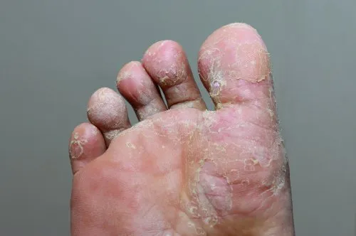
Fungal infections, or mycosis, are diseases caused by a fungus (yeast or mould). Fungal infections are most common on your skin or nails. Fungi are living things that are classified separately from plants or animals. They move around by spreading out or sending spores (reproductive parts) into the air or environment. Many fungi live naturally in our body, but can overgrow under certain circumstances.
A group of fungi that live off of skin, hair and nail cells (dermatophytes) cause ringworm. They can infect your feet (tinea pedis/athlete’s foot), your groin and inner thighs (tinea cruris/jock itch), your scalp (tinea capitis), your hands (tinea manuum), your facial hair and skin around it (tinea barbae) and other parts of your body (tinea corporis).
Many types of fungi cause infections of your fingernails or toenails (onychomycosis). This can cause discolored and cracked nails.
The fungus Malassezia causes skin discoloration called tinea versicolor or pityriasis versicolor.
A skin mycology test, also known as a fungal culture test, is a laboratory procedure used to identify and diagnose fungal infections of the skin, hair, and nails. It involves collecting samples from the affected area and analyzing them under a microscope or culturing them in a lab to identify the specific fungal species.
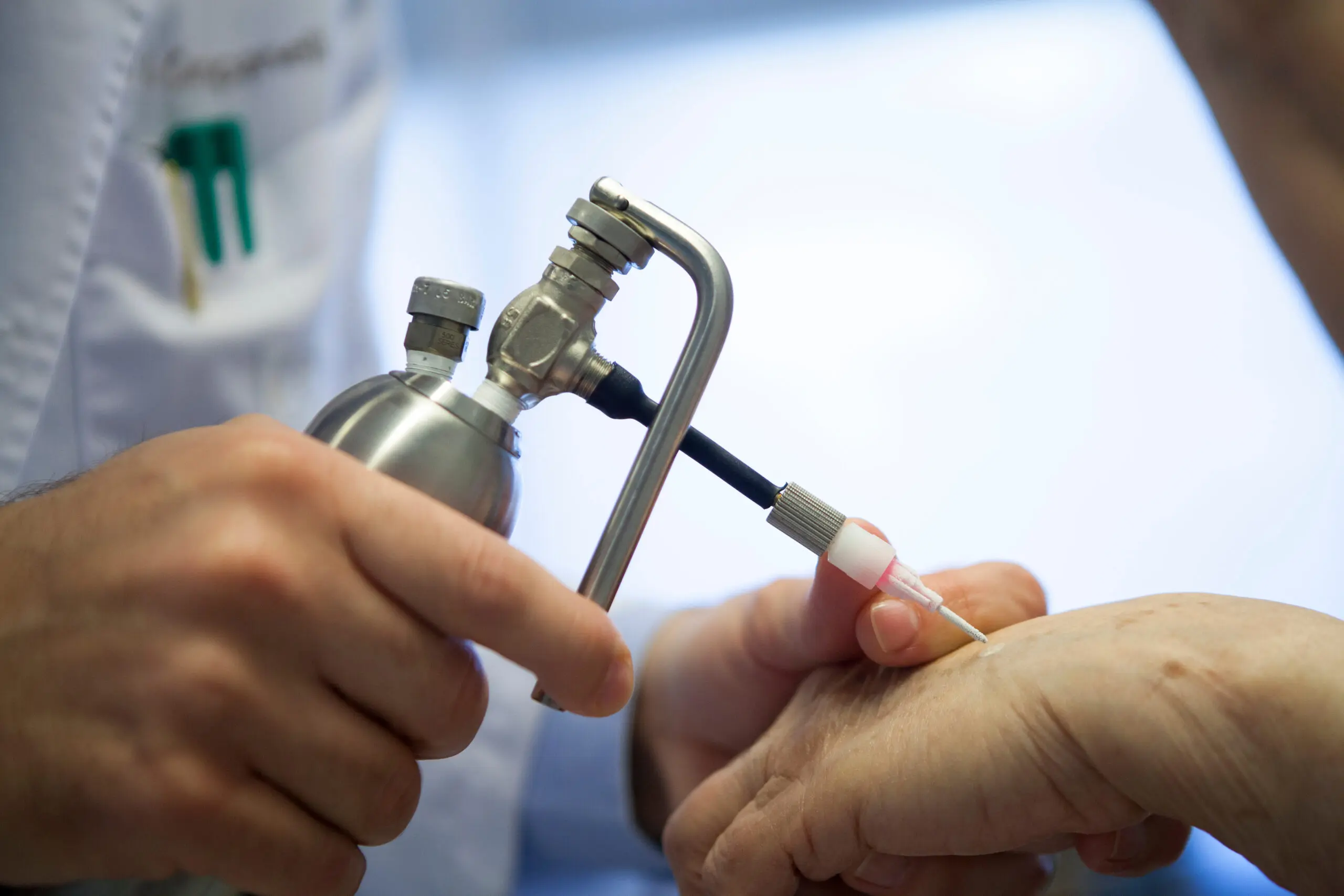
Cryotherapy is a method for treating some skin abnormalities has been in use for more than 150 years. The machine used to treat your skin is a product of modern technology that carefully controls a very cold liquid (nitrogen) such that it can be sprayed or touched onto any area of skin that needs treatment. The particular advantage of the treatment is that it replaces the need for a surgical operation. In effect the treatment is a carefully controlled burn.
Before the lesion is removed, the doctor will explain what they are going to do and will ask for permission (consent) to do the cryotherapy.
The procedure may simply cause a vague soreness or pain – this depends on the length of the freeze and the area being treated. After treatment marked redness always occurs together with some swelling. These changes usually last for a few days. In some people, particularly where the skin is rather thin and sensitive, a water (or blood) blister may form and fluid may discharge.
Once the fluid discharge or blistering stage is over a crust may form which will eventually drop off. The area will usually heal within a few weeks – whilst the area is healing you can wash the affected area. If you have been treated with a long freeze you may be prescribed a steroid cream to use. Apply it twice daily on any clean dressing – unless otherwise instructed. Small areas can be covered with an Elastoplast type dressing. If you have not received a prescription use clean Vaseline twice daily. If undue discomfort or pain occurs after the treatment then a simple pain relief remedy such as Paracetamol or Aspirin may be taken for 3-5 days.
Occasionally scars develop. Pigmented or dark skins may lose pigment resulting in persistent pink patches. Alternatively darker patches may develop. Near the eye, swelling occurs for a few days – worse in the morning.
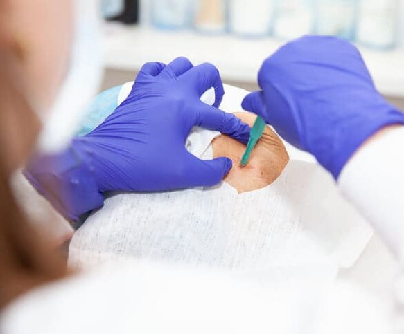
During a punch biopsy a small round plug of tissue is removed from the skin. The size of skin removed varies between 4-8mm. It is usually done to help provide a diagnosis rather than treat a condition.
Before the sample is removed, your doctor will explain what they are going to do and will ask for permission (consent) to do the biopsy. The procedure usually takes place lying down. A series of questions are asked out loud to make sure there will be no problems during the operation. This includes questions to check there are no allergies and that everyone confirms which lesion is being removed.
The skin is cleaned with an antiseptic liquid and a clean sheet or drape is put over the skin. The skin is made numb using an injection of local anaesthetic. This part can be uncomfortable but is very quick. The procedure itself should not be painful in any way. If it is, more local anaesthetic will be given. Generally, stitches are needed to close the wound and a plaster put on the wound.
The tissue that is removed is sent to the laboratory. A different doctor examines it under a microscope. They will write a report on what they find, but this can take up to 3-4 weeks.
The stitches are removed after 5 to 14 days, depending on where the surgery was. In Jersey General Hospital you will be given that appointment before you leave. You may have stitches that dissolve. These do not need to be removed.
The wound is not usually very sore afterwards. Any mild discomfort can be controlled with paracetamol.
Wound care advice will be given. Usually, the wound is covered and kept dry for 48 hours. After this time, the plaster is removed and brief showers are allowed (avoiding rubbing the wound with a towel after).
Increasing pain, redness or discharge is a sign of possible infection which would require medical attention.
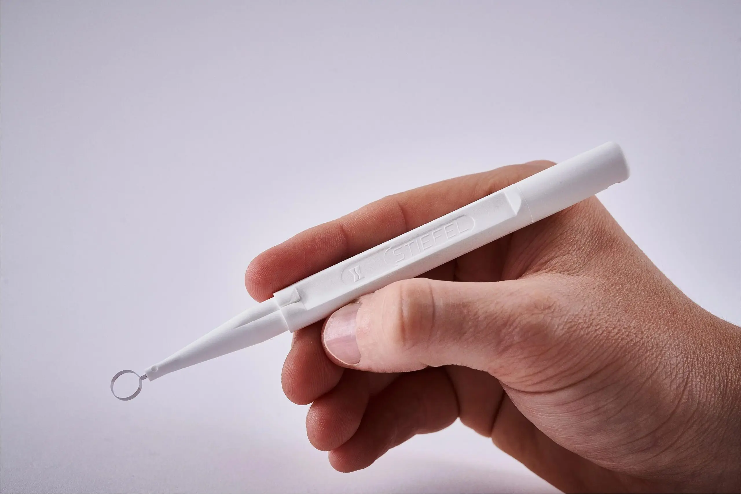
During a curettage and cautery a skin lesion is scraped off with a curette, which is like a small spoon with very sharp edges or a loop-like cutting tool.
Before the lesion is removed, the doctor will explain what they are going to do and will ask for permission (consent) to do the curettage and cautery. The procedure usually takes place lying down. A series of questions are asked out loud to make sure there will be no problems during the operation. This includes questions to check there are no allergies and that everyone confirms which lesion is being removed.
The skin is cleaned with an antiseptic liquid and a clean sheet or drape is put over the skin. The skin is made numb using an injection of local anaesthetic. This part can be uncomfortable but is very quick. The procedure itself should not be painful in any way. If it is, more local anaesthetic will be given.
The tissue that is removed is sent to the laboratory. A different doctor examines it under a microscope. They will write a report on what they find, but this can take up to 3-4 weeks.
Initially the wound looks like a deep graze or cigarette burn. This will gradually heal over to form a scar.
Wound care advice will be given. Usually, the wound is covered and kept dry for 48 hours. After this time, the plaster is removed and brief showers are allowed (avoiding rubbing the wound with a towel after).